Bye, Bye Dental Fillings: No ‘Fill And Drill’ Dentistry Technique Prevents Tooth Decay

Many of us fear to go to the dentist on the off chance the dentist will need to drill our teeth. However, a dental care technique devised by researchers at the University of Syndey could stop, reverse, and prevent tooth decay without the traditional “fill and drill” approach.

“It’s unnecessary for patients to have fillings because they’re not required in many cases of dental decay,” said Wendell Evans, lead author of the study and associate professor at the university, in the news release.

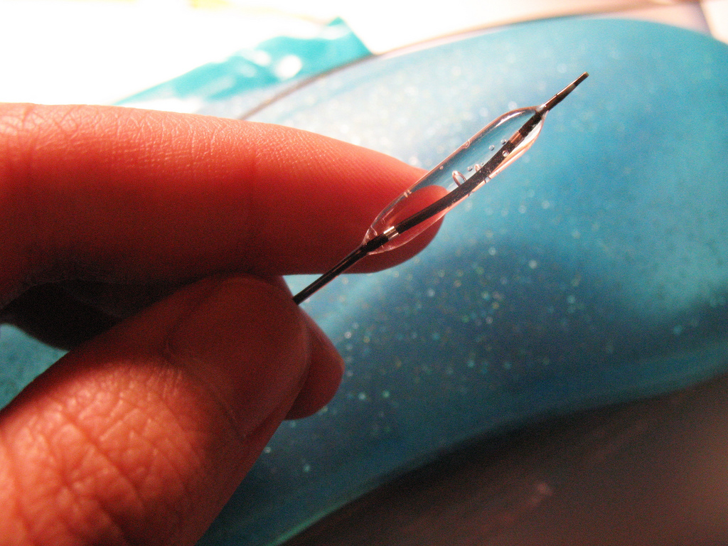
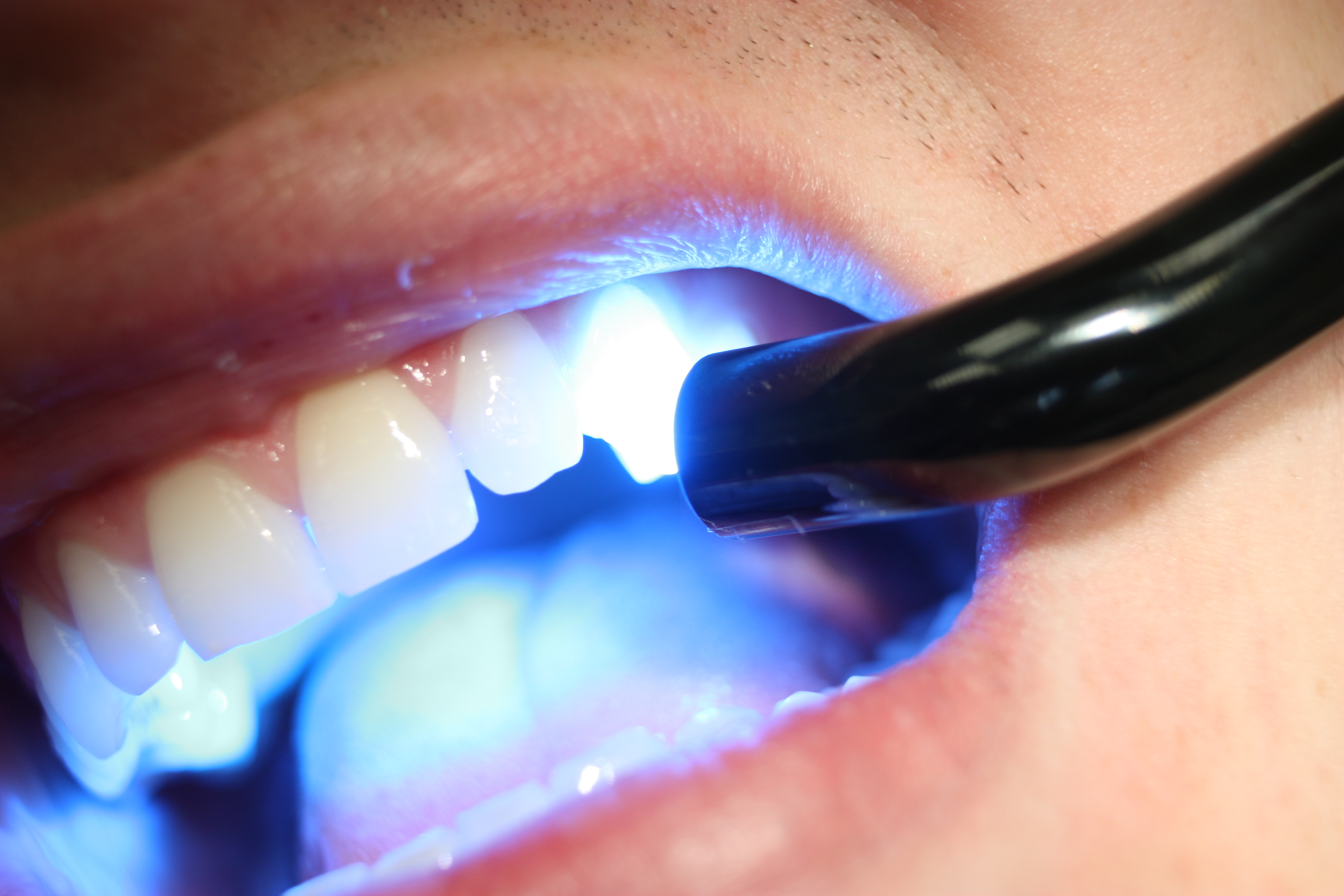
It was once believed that tooth decay was a rapidly progressive disease and the best way to manage it was to identify early decay and remove the tooth surface from breaking into cavities. After removing the decay, the affected tooth would then be restored with filling material, a process which would come to be known as “filling and drilling.” However, Evans explained, it takes four to eight years before tooth decay reaches the inner layer of a tooth. While tooth decay begins to become noticeable in the outer layer of the tooth or the enamel, the time window is sufficient to offer an alternative preventive treatment for the patient.

Enter Evans and his colleagues. They devised a process known as Caries Management System (CMS) technique, a set of protocols which cover the assessment of decay risk, the interpretation of dental X-rays, and specific treatment of early decay (decay that is not yet a cavity). The treatment involves four aspects: application of high concentration fluoride varnish to sites of early decay; attention to home tooth brushing skills; restriction of between-meal snacks and beverages containing added sugar; and risk-specific monitoring. The CMS technique was proven successful when first tested on high-risk patients at Westmead Hospital in Sydney.

“It showed that early decay could be stopped and reversed and that the need for drilling and filling was reduced dramatically. A tooth should only be drilled and filled where an actual hole in the tooth (cavity) is already evident,” Evans said.

To observe the effects of this technique on tooth decay, researchers assigned 19 patients at high risk for tooth decay to an experiment or control group for three years in general dental practices in New South Wales and Australian Capital Territory. Follow-up studies were conducted at two and four years after the clinical trial. After seven years, tooth decay reduced dramatically by 30 to 50 percent in patients in the CMS program compared to the control group.

However, the researchers stress there needs to be open communication between dentists and their patients to reduce decay risk and the need for fillings. “…Patients play an important role in their treatment. This treatment will need a partnership between dentists and patients to be most successful,” Evans said.

Patients can make the most of CMS by paying attention to their tooth-brushing skills at home and restricting between-meal snacks and beverages that contain added sugar. For example, when a tooth is exposed to acid frequently in foods or drinks that contain sugar and starches the acid build-up will cause the enamel to lose minerals, according to the National Institute of Dental and Craniofacial Research. A white spot can appear where minerals have been lost, which is a sign of tooth decay. The enamel can repair itself by using minerals from saliva, and fluoride from toothpaste or other sources.

The CMS technique could help the 92 percent of adults with tooth decay in their permanent teeth in the U.S. This suggests it is imperative to start adopting more oral care practices at home. The prevalence of untreated decay in permanent teeth is highest among adults between the ages of 20 to 34 years a population that has most of their teeth.

When it comes to tooth decay, prevention may be the best idea.

Source: Evans W et al. “No-drill” dentistry stops tooth decay. Community Dentistry and Oral Epidemiology. 2015.

By Lizette Borreli / Medical Daily

Helping Stroke Patients Using Software That Helps Identify Who Would Benefit Most From Risky Angioplasty Procedure

Angioplasty is a procedure that can help to reduce the risk of stroke recurrence by restoring reduced or cut off blood flow to the brain. The procedure is extremely risky, but recently researchers from the University of Illinois at Chicago may have found a way to ensure this operation is only conducted on patients who would benefit from it most.

A stroke occurs when blood flow to the brain becomes restricted or completely blocked. During angioplasty, these arteries in the brain are surgically reopened. This can help reduce the risk of patients suffering from a second stroke, but the science behind this procedure has not yet been perfected. Not only does it carry its own risks of harm, but it doesn’t always work in all patients. However, thanks to a new study now published in the online journal JAMA Neurology, this procedure may soon become a lot less risky.

For 22 months, the team followed 72 adult patients who had previously experienced a stroke or symptoms of a stroke in the back of the brain, known as the vertebrobasilar region. These patients also all had blockage to at least 50 percent of the arteries in the vertebrobasilar region.

A software program is known as Noninvasive Optimal Vessel Analysis (NOVA) was used to measure the blood flow to the back of the volunteers’ brains. According to the press release, NOVA software can quantify the volume, velocity, and direction of blood flowing through any major vessel in the brain using standard MRI equipment.

By the end of the study, about a fourth of the volunteers were found to have a diminished blood flow in the back of the brain. In addition, these patients had 12- and 24-month stroke-free survival rates of 78 percent and 70 percent, respectively. This was compared to 96 percent and 87 percent for patients who had normal blood flow. By the one-year mark, patients who had lower blood flow were five times more likely to have a second stroke than patients with higher blood flow to the back of the brain.

Based on these results, the team concluded that patients with diminished blood flow to the vertebrobasilar have significantly more to gain from undergoing this procedure. In addition, the team concluded that angioplasty may actually do more harm than good to patients without restricted blood flow in the vertebrobasilar area since other arteries seemed to be doing their job in ensuring that blood reaches that area, explained lead researcher Dr. Sepideh Amin-Hanjani in a statement.

Source: Amin-Hanjani S, Pandey DK, Rose-Finnell L, et al. Effect of Hemodynamics on Stroke Risk in Symptomatic Atherosclerotic Vertebrobasilar Occlusive Disease. JAMA Neurology. 2015.

By Dana Dovey / Medical Daily

‘Bone Foam’ That Gets Injected Into Bone Could Help Treat Osteoporosis And Other Bone Diseases

Our bones are constantly regrowing. They attain peak mass sometime in our 20s before slowly getting weaker as we age. This increases many people’s risk of osteoporosis, a condition characterized by weak and brittle bones, and their subsequent risk of fractures from something as minute as a cough. To combat this decline in bone strength, researchers from France have developed an injectable foam that helps to repair bones.

Injectable calcium phosphate cement (CPCs) are often used in orthopedics and traumatology. They’re made of bioactive and biodegradable grafting materials that come in powder and liquid form. When mixed together, they create a calcium-like material similar to the calcium in our bones. This mixture allows surgeons to fix and harden degenerative bones over the course of their surgery with relative ease and flexibility. It’s also biocompatible, self-setting, and non-toxic.

The new study, published in Acta Biomaterialia, builds on this material. Before, scientists had difficulty making CPCs macroporous or having pores larger than 50 micrometers. While studies have found conflicting evidence regarding how porous bones should be to facilitate regrowth, the range seems to be between 100 and 300 micrometers ? in people with osteoporosis, bones are far more porous.

Led by Pierre Weiss, from the University of Nantes, the research team developed a type of CPC that is both self-setting and macroporous. To do this, the researchers created a foaming agent that produces air bubbles in the CPC mixture. They loaded one syringe with monosodium phosphate, a common industrial chemical, and a second with silanized-hydroxypropyl methylcellulose, a type of hydrogel into another. Then, they connected the two. By plunging both syringes simultaneously, they were able to create a hydrogel foam, which is meant to create larger pores. They then inserted the foam into another syringe connected to a syringe with CPC paste. Again, they plunged both syringes, which then created the foamed CPC paste.

To determine if the foam could actually repair degenerated bone, the researchers applied it to tiny defects in the thigh bones of rabbits. After a week, they found the foamed CPC had entirely filled the defect as well as maintained the macroporous structure they were looking for. Six weeks later, they found it had acted as a scaffold to facilitate new bone growth; there were newly formed bones within the CPC itself, fitting together uniformly.

To the best of the team’s knowledge, this is the first time a hydrogel foam has been used in conjunction with CPCs to create a macroporous paste. The researchers also noted their CPC paste “exhibits a good injectability and an excellent cohesion, which are needed for clinical application.”

Further studies are needed to show just how effective their paste can be, but the researchers believe it will one day help to prevent some eight million osteoporosis-related fractures each year across the globe.

Source: Weiss P, et al. A simple and effective approach to prepare injectable macroporous calcium phosphate cement for bone repair: Syringe-foaming using a viscous hydrophilic polymeric solution. Acta Biomaterialia. 2015.

By Steve Smith / Medical Daily

Blood Test For Testicular Cancer Patients Can Tell Who Needs Chemotherapy, Predict Risk Of Relapse

Researchers have developed a new test capable of identifying which patients are at greatest risk of suffering a relapse from testicular cancer. The test, which involves assessing three features of a testicular cancer tumor, could be used to help clinicians decide on the proper form of follow-up care.

“Our research has led to the development of a test that can detect patients that will benefit from treatment up front and spare those who are at lower risk from the side effects of chemotherapy,” said Janet Shipley, professor of cancer molecular pathology at The Institute of Cancer Research, London, in a press release.

Even if there is no evidence of a tumor spreading, the test can give patients a score that tells them how likely they are to relapse within two years. It’s highly unusual to suffer a relapse outside of this time frame. Researchers tested levels of CXCL12, an antimicrobial gene; the percentage of the tumor showing cancer stem cells; and whether or not blood vessels were present in the tumor. After scoring 177 tumor samples, the team divided up the patients into three different risk groups.

The majority of the patients tested were in the low-risk group, where 94.3 percent of patients did not relapse within two years. The moderate risk group saw 65.9 percent of patients stay relapse-free within the same time, while the high-risk group only had a dismal 30 percent of patients remain relapse-free. The researchers validated the test with an additional 80 patients after the initial study.

Testicular germ cell tumors are the most common solid malignant tumor in young Caucasian men. Properly identifying who would benefit from chemotherapy is a huge step forward in the treatment of these tumors.

“Chemotherapy is extremely effective in treating testicular cancer, but it can have long-term consequences for a patient’s health and wellbeing,” Shipley said. “Patients deemed at low risk of relapse could simply be monitored, and potentially could avoid chemotherapy.”

Shipley said avoiding chemotherapy (if possible) is particularly important for cancers like this since they would affect young adults who would have to live with chemotherapy side effects for many years after treatment.

Robert Huddart, professor of urological cancer at the Institute of Cancer Research, London, and a consultant at The Royal Marsden NHS Foundation Trust, said patients with this kind of germ cell tumor have to make a difficult choice between being monitored or receiving chemotherapy to reduce the risk of relapse.

“Our study may help men make this decision as among the three groups of patients identified, we have found one with a very low risk of relapse where surveillance would seem the best choice, and a small group of men who have a very high risk of relapse, who could be targeted with chemotherapy,” he said. “We now need to test this prognostic index in larger groups of men in the clinic.”

Source: Gilbert D, Al-Saadi R, Thway K, Chandler I, Berney D, Shipley J, et al. Defining a New Prognostic index for Stage I Non-seminomatous Germ Cell Tumors using CXCL12 Expression and Proportion of Embyronal Carcinoma. Clinical Cancer Research. 2015.

By Ali Venosa / Medical Daily

Drug Toxicity Reduced Through New Liquid Salt Formula: Researchers Deliver Propranolol With Reduced Toxicity

Topical drugs are used for an array of conditions, but skin toxicity remains a major challenge in the design and use of new drug formulas. A team of researchers from the University of California, Santa Barbara (UCSB) came up with a possible solution to the problem, demonstrating a novel formulation of a drug as a liquid salt.

Topical drug toxicity often occurs because drugs have to be dissolved in organic solvents, many of which are toxic to the skin. Some drugs, such as propranolol, show dose-dependent skin toxicity on their own. Propranolol is often used to treat high blood pressure, chest pain, and uneven heartbeat.

The UCSB team demonstrated the drug as a liquid salt, which allowed delivery through the skin with reduced toxicity, it eliminated both possible sources of toxicity. The liquid salts are fluid, mitigating the need for organic solvents, and counterions used in the formation of the salts shielded the drug’s charge, which further reduced the drug’s toxicity.

“Propranolol is positively charged, which is a likely source of its toxicity,” explained Professor Samir Mitragoti, of the University of California, Santa Barbara, and lead author of the paper, in a press release. “Shielding of this charge by association with a counter species in the liquid salt reduces its toxicity. These findings are broadly applicable to many charged drugs.”

Past studies have demonstrated how liquid salts can improve drug transport through the skin, but this study is the first to report the design of liquid salts aimed at minimizing skin toxicity. Formulations like this one may be able to broaden the spectrum of drugs that can be safely and effectively administered through a transdermal patch.

“An ideal drug liquid salt would need to permeate through the skin as an associated ion pair. Eventually, however, the drug and the counter ion must dissociate in blood to preserve drug’s therapeutic efficacy,” said Michael Zakrewsky, the co-first author on the paper. “We show that these attributes can be balanced through careful selection of counterions.”

Source: Aoyagi K, Zakrewsky M, Mitagotri S. Formulating propranolol as an amorphous melt affords reduced skin irritation potential for transdermal drug delivery. Technology. 2015.

By Ali Venosa / Medical Daily